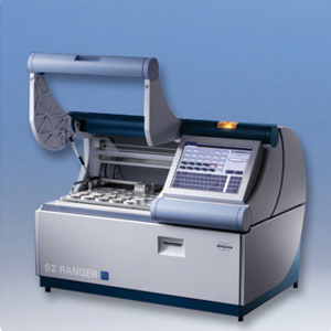
Description
Application:
- FESEM is used in the field of material science, geology, biological science, medical science and forensic science.
- It is used to obtain micrographs of surface morphology of the sample, compositional and bonding differences through contrast using BSE.
Principle:
Field Emission Scanning Electron Microscope uses a high energy electron beam emitted by an electron gun (tungsten wire) to create images of micro- and nano-sized samples by scanning the beam across the surface of the sample in a regular pattern. The electromagnetic lenses help in magnifying the object from 10 to 300,000 times. As the electron beam hits the sample surface, it interacts with the atoms on the sample and emits Secondary Electrons (SE – from top ~15 nm) and Back-Scattered Electrons (BSE – from top 40% of the sample). SE are low energy electrons
(<50eV) formed by inelastic scattering and are used to generate surface images.To increase the yield of SE, the samples are coated with ~10 nm of gold or platinum. BSE formed by elastic scattering is used to derive subsurface information.


Reviews
There are no reviews yet.